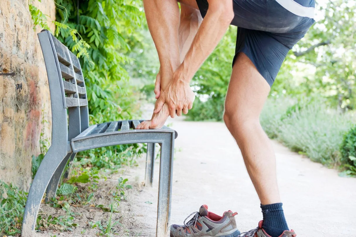
Your heel pain started few weeks ago. You assumed it was plantar fasciitis because it was common in runners, and that is what most runners assume. You stretched, you switched shoes, you tried a softer insole. The pain unfortunately does not ease. If anything, it has worsened with a “swollen feeling”. Walking is now uncomfortable. Standing for long periods produces a deep, dull ache across the entire heel rather than a sharp localised pain at the inside edge. You finally decide to see a doctor, get an X-ray, and the report comes back clear. No fracture visible, and report says, “no abnormalities detected”. The recommendation is to rest and stretch your calf. Two more weeks pass with no improvement, and you start thinking: maybe it is not plantar fasciitis at all.
This is the typical presentation of a calcaneal stress fracture we see in our clinic. The condition is regularly missed in its first 2 to 3 weeks because X-rays are unreliable in that window for most cases. Patients are often then treated for plantar fasciitis, heel fat pad syndrome, or general overuse heel pain, while the underlying microscopic crack in the heel bone is left unattended for too long and the recovery clock has not yet started. Recognising the pattern early changes the trajectory significantly.
A calcaneal stress fracture is a microscopic crack in the calcaneus, the heel bone, that develops from repetitive impact loading. It is different from a traumatic fracture, which results from a single high-energy event, usually a fall from height. The stress version develops gradually as the bone’s normal remodelling capacity is outpaced by the load being placed through it. The condition is most common in runners with sudden mileage increases, in military recruits during early training, in dancers and jumping athletes, and in older adults with reduced bone density. We often see this following Standard Chartered Marathon or Hyrox season.
Since calcaneal stress fracture often gets misdiagnosed as other heel pain conditions, to reccognise it earlier, it helps to know what its symptoms are like.
Symptoms of Calcaneal Stress Fracture
Stress fractures of the heel bone develop gradually. Therefore, symptoms often starts from having a mild heel discomfort, then becomes progressively more noticeable. The mild symptoms usually start after a higher-impact activity like a long-distance run.
- Deep, dull, aching pain across the whole heel – the discomfort is felt across the entire weight-bearing surface of the heel rather than at one specific tender point. Patients often struggle to localise it precisely.
- Pain that worsens with activity and persists with rest – unlike plantar fasciitis (which classically worsens on the first step after rest, then eases as you walk), stress fracture pain often gets worse with any weight-bearing activity and lingers as a deep ache even after stopping.
- Tenderness when squeezing the heel from both sides – one of the clinical tests we use, compressing the calcaneus between thumb and forefinger from the inner and outer sides of the heel reproduces the pain. This is the classic positive “squeeze test”.
- Mild swelling or “swollen feeling” at the heel – subtle puffiness around the heel can be present, but is not always obvious because it is mild.
- Pain that does not match the typical plantar fasciitis pattern – no clear morning first-step worsening, no specific tender point at the inside edge of the heel.
- Progressive worsening over weeks – rather than fluctuating or improving with rest, the pain tends to escalate steadily until the patient stops all activity entirely.
- In some cases, pain at night or with non-weight-bearing positions – typically indicates a more significant crack, so immediate medical attention is necessary.
Causes of Calcaneal Stress Fracture
Calcaneal stress fractures develop when the load placed through the heel bone exceeds its capacity to remodel and adapt. Multiple factors contribute, and most clinical cases involve a combination of triggers and pre-existing risk factors.
What Causes Calcaneal Stress Fracture?
- Sudden increase in running or impact training volume – the most common single trigger. A jump from 30 km per week to 60 km, a new running program, or rapid marathon training escalation.
- Repetitive jumping or landing activities – basketball, volleyball, jump rope training, and dance create repeated heel impact loading.
- Hard training surfaces – shifting from grass or turf to concrete or tiled floor increases the impact force the heel must absorb with every step.
- Poor shoe cushioning or barefoot training – worn-out running shoes, minimalist footwear, or barefoot training without proper transition, or training shoes used long past their intended mileage.
- Military training – the classic National servicemen context. New recruits in early phases of basic military training are at higher risk due to sudden high-volume marching, running on hard surfaces, especially in those hard boots.
- Poor recovery between training sessions – inadequate sleep or rest, suboptimal nutrition, and back-to-back hard training days reduce the body’s capacity to remodel bone effectively.
Who Carries a Higher Baseline Risk?
- Long-distance runners and high-performance athletes, particularly during marathon training cycles.
- NS recruits in the early phases of basic military training.
- Low energy availability or Relative Energy Deficiency in Sport (RED-S), which reduces bone density and remodelling capacity. For females, menstrual irregularity is a red flag.
- Adults with osteoporosis or reduced bone mineral density, including postmenopausal women not on hormone or bone-protective therapy.
- People with vitamin D deficiency, which is quite common in Asia due to sun avoidance.
- Older adults who suddenly increase walking or activity (for example, starting a new exercise routine without graduated progression).
- People with structural foot factors, such as high arches.
Need Help? See A Podiatrist Today
Conditions Commonly Mistaken for Calcaneal Stress Fracture
Most clinician would not consider a presenting heel pain case as calcaneal stress fracture immediately because they are far less common, and that is why it often get misdiagnosed. Besides, there are a few conditions that present similarly, especially in the beginning. Differentiating them matters because management is fundamentally different, and applying incorrect treatment can lead to much longer recovery.
- Plantar fasciitis – Definitely the most common misdiagnosis we see. The distinction lies in the pain pattern. Plantar fasciitis pain is typically sharp, localised to the inside edge of the heel or the arch. It is usually worst on the first few steps after rest and improves with walking. Calcaneal stress fracture pain is deeper and duller, spread across the whole heel, gets worse with continued activity, and lingers or throbs after stopping.
- Heel fat pad syndrome – Both conditions cause pain across the underside of the heel, and both worsen with hard surfaces and barefoot walking. The key difference is that fat pad syndrome often results in bruising pain that is at the fatty cushion layer, which feels less deep compared to calcaneal stress fracture. For heel fat pad syndrome, squeezing the fat pad reproduces the pain as compared to squeezing the sides of the heel bone for calcaneal stress fracture.
- Retrocalcaneal bursitis or insertional Achilles tendinitis – Less commonly misdiagnosed as, but still happens when clinicians did not perform a thorough check. The distinction is straightforward: pain at the back of the heel rather than underneath the heel, with tenderness directly at the Achilles tendon attachment.
Managing and Preventing Calcaneal Stress Fracture
When the diagnosis and underlying causes are clear, the management of a calcaneal stress fracture becomes fundamentally simple. The key principle is straightforward: the bone is injured and needs to be unloaded to allow it to remodel and heal. It is similar to treating any other bone fractures.
There is no advanced therapy, no orthotic, and no exercise protocol that bypasses this basic requirement. Continuing to load the bone while the crack is active risks the stress fracture progressing to a bigger fracture, which would then require a significantly longer recovery. This is why if a calcaneal stress fracture is being treated as plantar fasciitis or Achilles tendonitis, it usually gets worse, because part of treating those conditions involves a loading regimen.
To tackle the condition appropriately, the initial phase focuses on making sure the diagnosis is right and immediately removing the load from the bone:
- Clinical assessment to identify the diagnosis, including the squeeze test and palpation pattern.
- Referral for MRI may be necessary to confirm the diagnosis. X-rays are generally unreliable and unremarkable in up to 85% of cases, especially taken during the first 2 to 3 weeks of symptoms.
- Fitting of a CAM walking boot to protect the heel and redistribute load to the lower leg.
- Complete cessation of running, jumping, and high-impact loading for the duration of boot wear, typically 4 to 6 weeks. It should only be removed when showering or sleeping.
- Crutches may be required if weight-bearing remains painful even in the boot.
- Extracorporeal shockwave therapy and magnetotransduction therapy can be considered at this stage to accelerate the healing response and reduce pain and inflammation.
The next phase, typically between weeks 4 to 8, focuses on protected healing:
- Transition out of the walking boot if possible. If pain has significantly reduced, a transition to a supportive, high-cushioning footwear should be made. Footwear should be worn both indoors and outdoors.
- For cases associated with foot structural issues like high arches, a custom insole may be considered to reduce heel impact during activity
- Begin body reconditioning by switching to non-impact cardiovascular fitness through swimming, cycling on stationary equipment, or other activity that minimises weight-bearing.
- Vitamin D, calcium, and overall nutrition optimisation to support bone remodelling.
- Assessment of contributing factors, including reviewing training methods, and addressing any underlying issues like RED-S or osteoporosis.
The return phase, typically weeks 8 to 16, focuses on a gradual return to activity:
- Start a progressive weight-bearing plan, from walking, to incline walking, then to slow jogging.
- Build running volume back gradually using a structured return-to-running program. Returning to pre-injury mileage can take up between 3 to 6 months, so patience is key.
- If major muscular decondition has occurred due to a period of rest, seeing a physiotherapist for a structured strengthening program will be highly beneficial
For ongoing prevention after recovery:
- Follow the 10% rule for training load increases to avoid a sudden increase in activity.
- Mix training surfaces and avoid sustained running on concrete or pavement.
- Rotate between two or more pairs of running shoes, and always replace worn-out running shoes in a timely manner.
- Maintain bone health through adequate nutrition.
- Take sufficient rest and recovery days to allow the body to heal and the bones to remodel.
- Where biomechanical factors contributed, use the prescribed custom orthotics during sustained training.
Surgery is very rare in calcaneal stress fracture management and is reserved for non-healing fractures that have not responded to conservative care over many months. Most cases should heal completely with appropriate offloading and time.
Have Your Calcaneal Stress Fracture Managed at Straits Podiatry
Calcaneal stress fracture is a condition where early recognition makes a meaningful difference. Patients who arrive with several weeks of escalating heel pain after a recent training change, where usual heel pain treatment advice has not helped, where X-ray results is too reassuring, are exactly the patients in whom this condition is often hiding. At Straits Podiatry, an assessment for suspected calcaneal stress fracture focuses on the patient’s activity history, the clinical examination (squeeze test, palpation, weight-bearing reproduction), and a clear discussion of whether MRI confirmation is needed and how to arrange it.
From there, a management plan is built around your specific case. This may include a comprehensive lower-limb assessment to identify any contributing biomechanical and training factors, fitting and guidance on walking-boot use, custom foot orthotics, and structured rehabilitation to address deconditioning and return to sport.
When heel pain starts to get in the way of your training, you should take care of it before it gets worse. Speak with our team or book a consultation for an assessment and our podiatrists will assist you.
Frequently Asked Questions About Calcaneal Stress Fracture
How long does a calcaneal stress fracture take to heal?
A calcaneal stress fracture usually takes between 4 to 6 weeks to heal, but can take months before you can return to sport. However, the duration is dependent on appropriate management. At the start, protected weight-bearing in a walking boot is required for 4 to 6 weeks, sometimes longer in cases with delayed diagnosis or more significant cracks. Full return to sport, especially back to pre-injury training volume, typically takes 3 to 6 months from the start of appropriate treatment.
Can I still walk with a calcaneal stress fracture?
Yes, but only with appropriate protection. The standard treatment is a walking boot (CAM boot) for daily activities, removed only for sleep and bathing. The boot redistributes load away from the heel and allows controlled weight-bearing without aggravating the lesion. Walking without protection during the healing phase risks fracture progression and significantly extends recovery. When pain settles after 4 to 6 weeks in a walking boot, you may transit into a pair of supportive, high-cushioning sports shoes for continued protection.
Why was my X-ray clear if I have a calcaneal stress fracture?
We get the question all the time, and the answer is that sometime the fracture is too small to detect on radiographs. Stress fractures are fine disruption of the soft bone rather than a clear break, so typically do not show on plain X-ray, especially during the first 2 to 3 weeks of symptoms. They only become more detectable when the fracture healing process has started, showing up as a sclerotic or bright line on X-ray. MRI is the gold standard for diagnosis because it can detect the bone marrow oedema and trabecular changes that X-ray could not.
How is a calcaneal stress fracture different from plantar fasciitis?
Plantar fasciitis pain is sharp, usually localised to the inner edge of the heel where the plantar fascia attaches, and worst on the first few steps after rest. Calcaneal stress fracture pain tends to be duller and deeper, and spread across the whole heel. Calcaneal stress fracture also worsens with continued activity, and lingers as a dull ache after stopping.
When can I return to running after a calcaneal stress fracture?
Return to running following a calcaneal stress fracture must be gradual and with a structured plan. Our best advice is to start once you are comfortable walking on a treadmill, especially on a incline, without any symptoms. Following that, you can begin slow jog, keeping the distance short (2 to 3km) to begin with, and increase 10% each week. Patience is very important to avoid setbacks. Returning too early is the single most common reason for re-injury, and the consequences (potential complete fracture) are significant enough that the conservative approach is the right one.
Share this with someone you know