

You sprained your ankle weeks ago and it mostly settled, except for a deep ache inside the joint that will not go away. It flares with prolonged walking or sport, and sometimes the ankle clicks, catches, or even locks. This is the pattern of a talar dome injury: damage to the cartilage and underlying bone on the upper surface of the talus, the bone that sits inside the ankle joint. It most often develops after an ankle sprain, when a forceful twist compresses the talus against the shinbone hard enough to bruise or fracture the cartilage surface.
Because the symptoms closely mimic a slow-healing sprain, talar dome injuries are commonly missed in the first round of assessment. Left undiagnosed, the damaged cartilage can fail to heal properly, leading to chronic pain, joint inflammation, and an elevated risk of early ankle arthritis. Identifying these injuries early changes the recovery pathway significantly.
Symptoms of Talar Dome Injuries
Talar dome injuries often hide behind the symptoms of the original sprain, so the clue is usually in what does not settle over time. The pattern to watch for:
- Deep ache inside the ankle joint: a pain felt deep within the joint rather than over the surface, and most patients find it hard to pin-point the exact location.
- Pain and swelling that flares with load: discomfort and puffiness that usually build with walking, running, or pivoting, then ease with rest.
- Pain that does not reproduce with ankle joint movement: unlike an ankle sprain, moving the ankle from inversion to eversion does not easily reproduce the pain.
- Difficulty bearing weight, especially on uneven ground: for severe cases, the pain tends to increase on rough or sloping surfaces.
- Stiffness and reduced range of motion: the ankle may not bend or point as freely as the other side, often most noticeable first thing in the morning or after a period of sitting.
- Clicking, catching, or locking: a mechanical catch or lock during movement, which can point to a cartilage flap or a loose fragment moving within the joint.
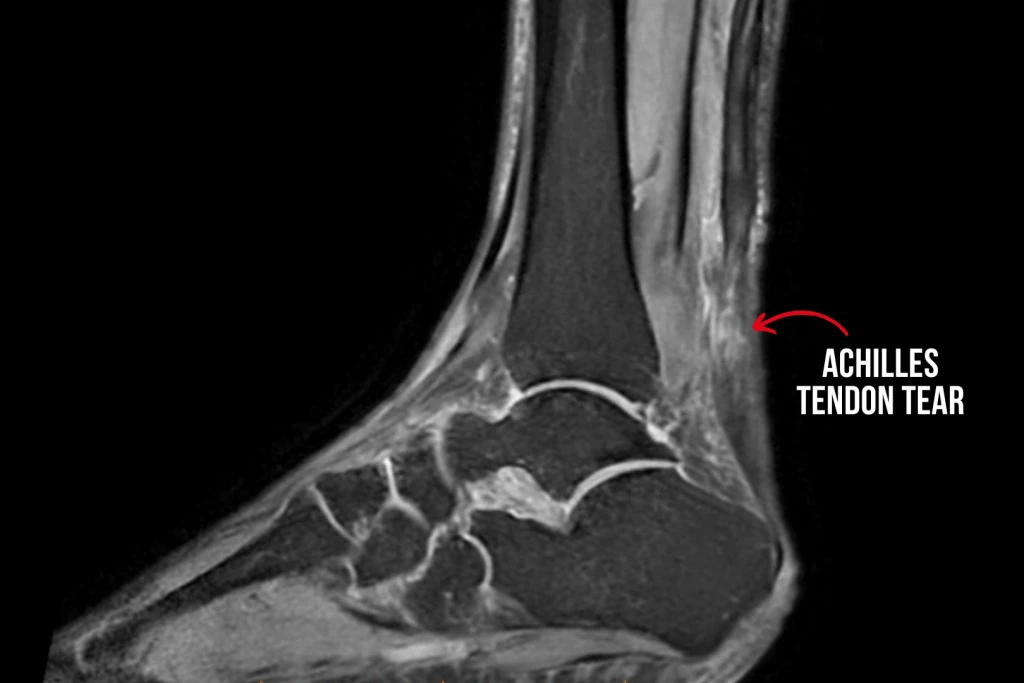
Types of Talar Dome Injuries
Talar dome lesions are usually graded by depth and stability of the cartilage and underlying bone. The grading matters for the treatment plan, and mismanagement can lead to a much longer recovery.
Grade 1
The cartilage is softened but the surface stays intact. This is the mildest form, where the injury is confined to the cartilage without a visible break. Because nothing has fractured, it can be the hardest grade to see on imaging, and it is the stage most likely to settle with offloading and protected loading.
Grade 2
There is a partial cartilage fracture, but the surface fragment stays attached to the underlying bone. The cartilage has started to break, yet the joint surface is still largely held together. Symptoms tend to be more persistent than a Grade 1, and healing depends on keeping load off the area while the cartilage stabilises.
Grade 3
The cartilage tears through its full thickness and the underlying bone is exposed, but the fragment has not yet moved. Because the break now reaches down to the bone, this grade is more likely to ache or catch under load. It usually needs closer monitoring, since it can progress if the fragment loosens.
Grade 4
A fragment detaches and floats loose within the joint. The piece of cartilage and bone has separated completely and can move around inside the ankle, which is what produces the clicking, catching, or locking. This is the most advanced grade and the one most likely to need surgical removal or fixation of the loose fragment.
Lower grades typically respond well to conservative care. Higher grades, especially Grade 4, often need surgical intervention.
Causes of Talar Dome Injuries
The most common cause is an acute ankle sprain, particularly a high-energy twist or a sprain combined with a vertical load (such as landing from a jump). The talus compresses against the tibia and damages the cartilage. Other contributors include direct trauma to the ankle and repetitive impact in sports such as basketball, football, or running. Recurrent ankle sprains are a common driver, as seen in most of our patients. Foot posture matters too. A high arch and severe flat feet (especially with hypermobility) can cause excessive, unstable ankle and subtalar joint movements. Underlying joint conditions such as early arthritis or osteochondritis can also predispose to talar lesions.
Who Carries a Higher Baseline Risk?
- People with a history of moderate or severe ankle sprains.
- Athletes in high-impact, pivoting sports.
- Those with chronic ankle instability or recurrent sprains.
- People with high-arched or significantly pronated foot postures.
- Patients with previous ankle fractures or established ankle arthritis.
Conditions Commonly Mistaken for Talar Dome Injuries
Most patients report that their problem has been treated as a slow-healing ankle sprain, until they realise the continued rehab is not working. The deep, persistent ankle pain of a talar dome injury can resemble several other post-sprain problems, and because plain X-rays do not always show cartilage damage clearly, an MRI is often needed to confirm the diagnosis when symptoms outlast the expected sprain recovery window.
Ankle impingement
Ankle impingement is pain caused by soft tissue or bony structures being pinched at the front or back of the ankle during end-range movement. The pain is usually provoked at the extremes of pointing or flexing the foot and is felt closer to the joint line, rather than as the deep ache within the joint that marks a talar dome injury. A talar dome injury also tends to flare with load and can click, catch, or lock when a cartilage fragment moves, which is not the impingement pattern.
Peroneal tendon problems
Peroneal tendon problems involve the tendons that run behind the outer ankle bone on their way to the foot. The pain follows the tendon line along the outside of the ankle and worsens with active eversion of the foot against resistance. Talar dome pain sits deep inside the joint and does not reproduce with that tendon-loading movement, which is the main way the two are told apart.
Chronic ankle instability
Chronic ankle instability is a recurring sense of the ankle giving way after previous sprains, driven by stretched ligaments and reduced proprioception. The dominant complaint is repeated rolling and a feeling of unsteadiness on uneven ground, rather than a fixed deep joint ache. The two often coexist, because the repeated sprains that come with instability are also a common driver of talar dome damage.
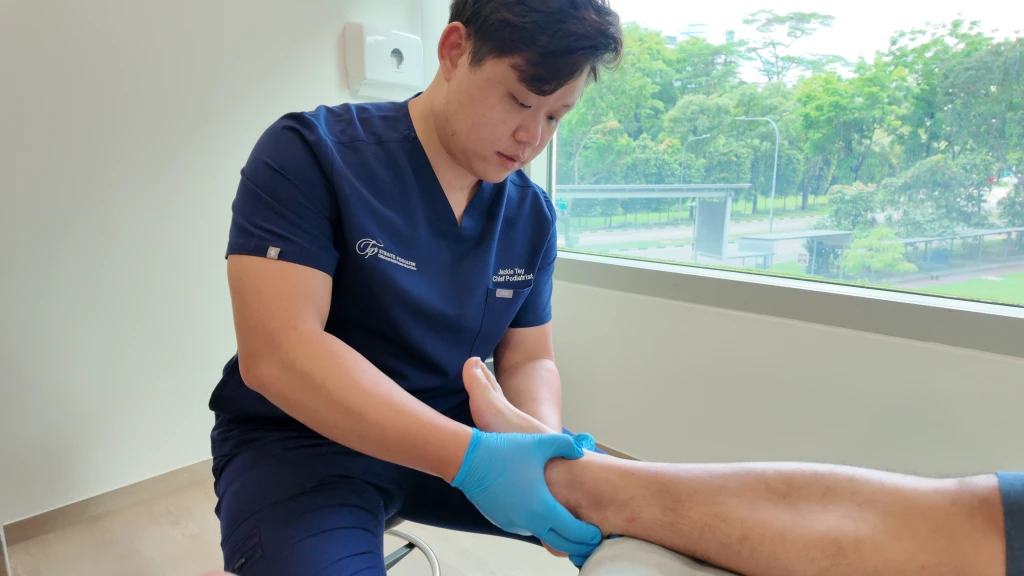
Treating and Preventing Talar Dome Injuries
Talar dome injury treatment is strongly guided by the lesion grade and the patient’s activity demands, because cartilage heals slowly and does not regenerate as readily as a tendon. Conservative care is the starting point for most low-grade lesions: protect and offload the cartilage early while strength and mechanics are restored, so the joint is not overloaded during healing. For higher-grade lesions and those with loose fragments, referral for surgical review is often necessary, though addressing the biomechanics and the rehabilitation that follows any procedure still matters. In some cases MRI does not fully show the extent of the damage, so an exploratory arthroscopic assessment may be needed.
Conservative treatment
Conservative care is built around offloading the cartilage, restoring joint function, and correcting the mechanics that were loading the lesion.
- Immobilisation and offloading: A period of immobilisation in a brace, walking boot, or cast allows the cartilage to settle without being repeatedly compressed. This is usually the first step for low-grade lesions that were detected early.
- Structured physical therapy: Structured physical therapy restores ankle strength and joint motion once the acute phase settles. This usually includes the balance and proprioception work that reduces the risk of a further sprain.
- Focused shockwave therapy: In mild to moderate cases, shockwave therapy can reduce pain and inflammation and stimulate a regenerative response around the lesion. Shockwave after surgery can also help accelerate recovery.
- Extracorporeal magnetotransduction therapy (EMTT): EMTT can be used alongside shockwave as a bone-healing option, which is useful where the lesion involves the underlying bone or bone marrow oedema.
- Custom orthotics or ankle-foot orthosis: Custom orthotics or an ankle-foot orthosis are used when foot mechanics are a driver of the lesion. The objective is to provide stability and reduce joint compression.
- Anti-inflammatory medication: Short courses of anti-inflammatory medication on medical advice may help settle pain and swelling during flare-ups.
When conservative care isn’t enough
Higher-grade lesions, loose fragments floating within the joint, and mild-to-moderate cases that continue to cause locking, swelling, or pain despite a structured period of conservative care typically need orthopaedic input, because the joint environment does not naturally regenerate cartilage well. Surgical options typically range from arthroscopic debridement and microfracture to cartilage transplantation or repair procedures, decided by a foot and ankle surgeon based on imaging. The podiatry role continues through the rehabilitation that follows any surgical procedure.
Preventing Talar Dome Injuries
Because most talar dome injuries follow an ankle sprain, prevention comes down to protecting the ankle:
- Rehabilitate each ankle sprain thoroughly the first time rather than returning to activity early.
- Avoid returning to sport before the joint has fully recovered.
- Wear footwear suited to high-impact sport, and address foot mechanics where a high-arched or flat foot posture is loading the joint unevenly.
Have Your Talar Dome Injury Managed at Straits Podiatry
Straits Podiatry assesses persistent post-sprain ankle pain with a structured clinical examination, gait analysis, and imaging where indicated. If a talar dome injury is suspected, your plan may include immobilisation, custom orthotics, ankle bracing, focused shockwave therapy, and structured rehabilitation through our physiotherapist. For higher-grade lesions, our podiatrists coordinate with orthopaedic surgical colleagues for further imaging and joint-preservation procedures.
Speak with our podiatrist in Singapore today, or book a consultation for an assessment and a tailored approach to manage your talar dome injury.
Frequently Asked Questions About Talar Dome Injuries
How do I know if my ankle sprain has caused a talar dome injury?
The biggest red flag is deep ankle pain that persists beyond the usual sprain recovery window of six to eight weeks, often with clicking, catching, or a sense of something inside the joint. If your sprain seemed to mostly heal but is now stuck at a plateau, or if you are getting recurrent ankle swelling without re-injury, it is worth getting the ankle reassessed by a podiatrist or orthopaedic surgeon.
Will a talar dome injury heal on its own?
Low-grade lesions can heal with rest, immobilisation, and load management, although the process is usually slower than ligament healing and the cartilage often does not fully restore to its original state. Higher-grade lesions, particularly those with loose fragments, generally do not heal without surgical intervention because the joint environment does not naturally regenerate cartilage well.
Do I need an MRI or an X-ray to check for a talar dome injury?
In clinic, you often need both. An X-ray shows bone but does not visualise cartilage directly, which means small to moderate cartilage tears can be missed. Even so, an X-ray is usually first-line to help rule out other bone and joint injuries. An MRI as a follow-up then shows the extent of the cartilage damage, bone marrow oedema (a sign of stress within the bone), and the surrounding ligaments, and confirms the diagnosis. Overall, MRI is generally the imaging of choice when a talar dome injury is strongly suspected. Even then, MRI cannot always show the full extent of the injury, in which case an exploratory arthroscopy may be needed.
Can I still play sport with a talar dome injury?
It depends on the grade, your pain level, and the treatment or support you have been given. Returning to sport too early risks worsening the cartilage damage and accelerating the development of ankle arthritis. Most patients need a period of offloading, orthotic support, structured rehabilitation, and a graded return to play. The exact timeline depends on the lesion grade and the demands of the sport, so this is best decided after imaging and clinical assessment.
Does a talar dome injury need surgery?
Whether surgery is needed depends on the lesion grade, the size of the cartilage defect, and how the ankle responds to a structured period of conservative care. Low-grade lesions without loose fragments are usually managed non-surgically with offloading, supportive footwear or orthoses, and progressive rehabilitation to restore ankle strength and proprioception. Higher-grade lesions, lesions with loose fragments inside the joint, and lesions that continue to cause locking, swelling, or pain despite conservative care typically need a surgical opinion. Surgical options range from arthroscopic debridement to microfracture and cartilage repair techniques, decided by a foot and ankle surgeon based on imaging.