
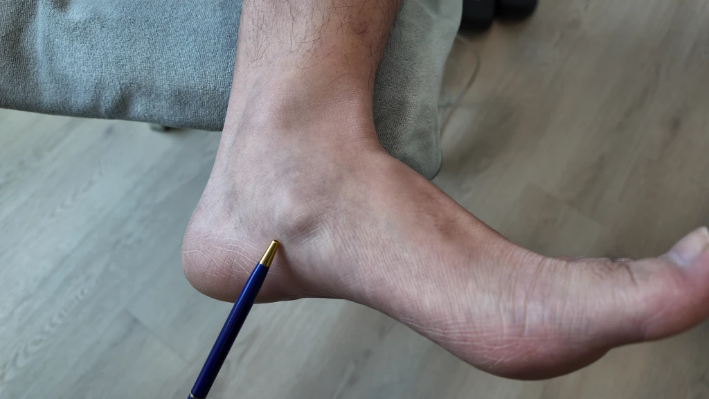
You notice a tender bony bump on the inside of the foot, just above the arch. Pressing on it reproduces a sharp pain. Long walks, dance class, or sports leave the area aching, and a pair of tight-fitting shoes can rub directly on the bump. For some, the pain has been there since adolescence. For others, it appears suddenly after an ankle sprain or a sudden increase in activity. The arch may also feel like it is collapsing during weight-bearing.
Os Navicular Syndrome, also known as an accessory navicular, develops when an extra bone or piece of cartilage near the inner side of the navicular becomes irritated. About one in ten people have this accessory bone, and most never develop symptoms. When pain does appear, it usually reflects strain on the posterior tibial tendon pulling on the accessory navicular, often combined with a flat foot posture. It is frequently misdiagnosed as posterior tibial tendonitis, and the two can coexist.
Symptoms of Os Navicular Syndrome
The symptom pattern is typically specific to the inner arch area. Pain location, tenderness on direct pressure, and a visible bony prominence usually point to the diagnosis.
- A tender bony lump on the inner side of the foot, just above the arch.
- Pain along the inside of the arch that worsens with activity or prolonged standing.
- Redness, swelling, and tenderness over the prominence, particularly after exercise.
- Discomfort with tight or stiff footwear that presses directly on the area.
- A feeling that the inner arch is collapsing when standing.
- Stiffness or difficulty pushing off during walking or running.
Types of Os Navicular Syndrome
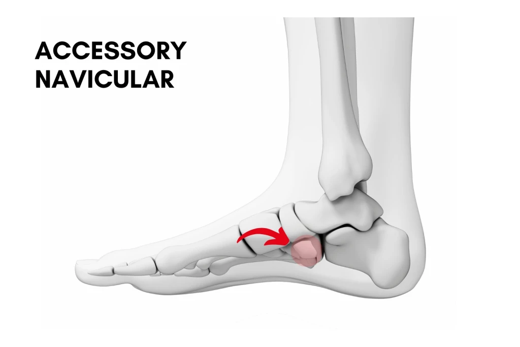
The accessory navicular is grouped into three types based on how the extra bone connects to the main navicular. The type influences how symptoms develop and how they are managed.
Type I
A small, separate ossicle (os tibiale externum) sits within the posterior tibial tendon, without a direct connection to the navicular. It is usually the least symptomatic type and is often found by chance.
Type II
The accessory bone is joined to the navicular by a fibrocartilage bridge (a synchondrosis). This junction can be injured or inflamed when the posterior tibial tendon pulls across it, which is why Type II is the type most often responsible for os navicular syndrome.
Type III (cornuate navicular)
The accessory bone has fused to the navicular, forming a prominent bony horn on the inner arch. Symptoms here tend to come from the prominence rubbing against footwear rather than from the junction itself.
Causes of Os Navicular Syndrome
The accessory navicular itself is a normal variant and is congenital, meaning you are born with it. The syndrome, which is the painful condition, develops when that extra bone becomes irritated. Most cases involve a combination of mechanical overload and a foot posture that puts the area under chronic strain.
What Causes Os Navicular Syndrome?
- Overuse from high-impact activity such as running, dancing, or jumping sports.
- Foot posture issues, particularly flat feet or overpronation, which stretch the posterior tibial tendon and increase the pull on the accessory navicular.
- Direct trauma to the inner arch from a fall or impact.
- Ankle sprains, where ankle inversion compresses the navicular bone against the medial malleolus (the inner ankle bone).
- Footwear that presses directly on the bony prominence, most commonly ski boots or ice-skating boots.
Who Carries a Higher Baseline Risk?
- Adolescents and young adults with active sport participation.
- People with flat feet or overpronation.
- Dancers, runners, and netball or basketball players.
- People with a family history of accessory navicular.
- Patients with a history of inner ankle injury.
Treating and Preventing Os Navicular Syndrome
Treatment focuses on settling the irritation around the accessory navicular and reducing the mechanical pull on it. Most cases do respond to conservative care, particularly when foot posture is addressed alongside the local symptoms. Surgery is reserved for symptoms that persist despite a fair trial of conservative treatment, or when severe arthritic changes are detected at the affected area.
Conservative treatment
Most cases settle with a combination of offloading the bone, calming the irritation, and rebuilding load tolerance.
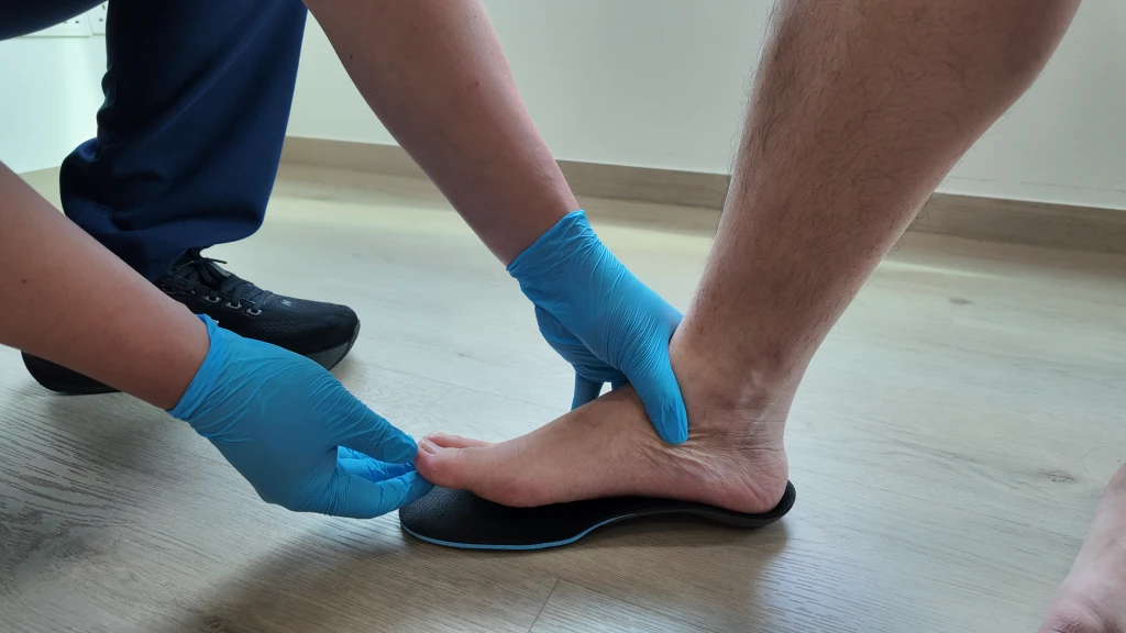
- Custom foot orthoses: Support the arch, control overpronation, and reduce the posterior tibial tendon pull on the accessory navicular. This is the mainstay where a flat or overpronating foot keeps loading the bone.
- Footwear guidance: A firm heel counter and adequate arch support help, while shoes that press directly on the prominence should be avoided. For ski or ice-skating boots, consider modifications to accommodate the prominence.
- Activity modification and relative rest: Swap high-impact sport for low-impact options such as swimming or cycling during a flare, and rest from the triggering activity while it settles. Ice after symptomatic sessions, and short courses of anti-inflammatory medication on medical advice, help calm the area.
- Extracorporeal shockwave therapy: Reduces pain and inflammation and supports tissue healing where the irritation is stubborn.
- Extracorporeal magnetotransduction therapy (EMTT): Used for its bone-healing properties and pain relief, which is helpful where the bony junction (a Type II synchondrosis) is irritated.
- Strengthening and rehabilitation: Strengthening the posterior tibial tendon, gluteal, and intrinsic foot muscles, along with calf stretching where tightness contributes, builds tolerance. A graded return to sport then rebuilds load progressively and helps prevent recurrence.
When conservative care isn’t enough
For persistent or severe cases, or where significant arthritic change has developed at the joint, surgery is considered. The options include excision of the accessory navicular and reattachment of the posterior tibial tendon, sometimes combined with structural correction of the underlying flat foot. Where surgery is indicated, a referral to an orthopaedic surgeon is arranged.
Have Your Os Navicular Syndrome Managed at Straits Podiatry
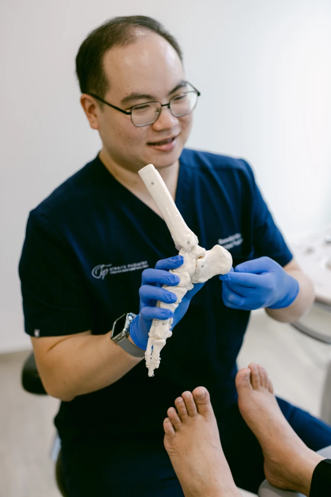
At Straits Podiatry, a suspected os navicular syndrome case will undergo an inner arch clinical examination and gait analysis to understand how foot posture and the posterior tibial tendon are loading the accessory navicular. From there, a management plan typically includes customised foot orthoses, footwear advice, and extracorporeal shockwave therapy where appropriate, alongside a graded loading programme.
Speak with our team or book a consultation for an assessment by our podiatrists in Singapore and a tailored approach to manage your Os Navicular Syndrome.
Frequently Asked Questions About Os Navicular Syndrome
Will I need an X-ray to diagnose this?
X-rays are commonly used to confirm the presence of an accessory navicular and to characterise its size and shape. Three main types exist, and the type influences how the bone connects to the navicular and how it presents clinically. A careful clinical examination is usually enough to point to the diagnosis, but imaging is helpful both to confirm the accessory bone and to rule out other causes of inner foot pain, such as a navicular stress fracture or posterior tibial tendonitis. MRI is occasionally needed for more complex presentations.
Can I still play sport with this condition?
Most patients return to sport once the area has settled and the underlying biomechanical factors are managed. During an active flare, switching to lower-impact activity (swimming, cycling, elliptical) protects the area while keeping cardiovascular fitness up. A staged return to running, dance, or court sports is then built, usually with continued use of orthoses and supportive footwear. Stopping sport entirely is rarely necessary unless symptoms are severe or surgery is being planned. Your podiatrist can help provide the right guidance for a return to activity.
Is the accessory navicular the same as posterior tibial tendonitis?
They are related, almost always coexist, but not the same. The posterior tibial tendon attaches to the navicular, and when an accessory navicular is present, the tendon often pulls on the accessory bone. Posterior tibial tendonitis is inflammation or degeneration of the tendon itself, while Os Navicular Syndrome involves pain at the accessory bone, often from that same tendon pull. The two frequently coexist, particularly in flat-footed patients. An accurate diagnosis matters because treatment overlaps but is not identical.
When is surgery needed?
Surgery is considered when symptoms persist despite a fair trial of conservative care, when pain significantly limits daily activity or sport, or when there is persistent swelling and a visibly prominent bump that does not settle. The most common procedure is excision of the accessory navicular with reattachment of the posterior tibial tendon. Where significant flat foot is contributing, a structural correction may also be added. If a flat foot remains after the procedure, we highly recommend seeing a podiatrist for custom foot orthosis support to reduce the load on the posterior tibial tendon and navicular bone.
Will the pain come back after treatment?
Recurrence depends on what is driving the symptoms. If the underlying flat foot or sport overload is not addressed, the same pull on the accessory navicular returns when activity ramps up again. Continued use of orthoses, supportive footwear, and ongoing strengthening of the posterior tibial tendon and gluteal muscles reduces the risk of recurrence considerably. Patients who treat the symptoms in isolation and then return to the same load pattern often see the pain return. A long-view plan is more durable.