
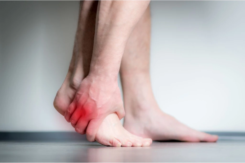
You had an ankle sprain six to eight weeks ago. The ankle has settled but the outside of your foot has not. Push-off hurts. Going up stairs hurts. Walking on the flat is fine for the first 10 minutes, then the lateral foot starts to ache. The standard ankle sprain rehab you finished did not change the pain on the outside of the foot, and the pain itself has shifted somewhere below the ankle. If this sounds like you, the culprit is likely cuboid syndrome.
The cuboid is a small wedge-shaped bone on the outside of the foot, sitting between the heel bone and the smaller toes. It is held in position partly by the peroneus longus tendon, which wraps around its underside on the way to the inside of the foot. During an inversion ankle sprain, it triggers the tendon reflex to resist the rolling motion and can pull the cuboid slightly out of its normal alignment, leaving it stuck in a partially displaced position long after the original ankle injury has healed. The condition is often missed because the pain has moved away from the ankle and standard ankle sprain rehab does not address it. Identifying it accurately prevents you from going through many unnecessary treatments for months.
Symptoms of Cuboid Syndrome
The pain pattern of cuboid syndrome is not that unique because a few other conditions can produce similar pain, particularly when there is a recent ankle sprain history. Location combined with timing are the most reliable early differentiator from residual sprain trouble.
- Pain localised to the lateral midfoot: the most tender spot sits around 1 finger below the dimple at the ankle and closer to the outer half of the foot.
- Pain that worsens with push-off, stairs, and jogging: anything that loads the lateral column of the foot reproduces or aggravates the pain. Walking on the flat is often tolerated for the first few minutes, then the ache builds.
- Occasional mild lateral arch ache: patients sometimes describe a vague ache along the outer arch, particularly after long days on their feet.
- Reduced shock-absorption sensation on the affected side: the foot feels harder, less springy, particularly during running.
- Pain reproduced by compression of the cuboid: pressing the cuboid between a thumb on the top of the foot and a finger underneath reproduces the patient’s pain. This is the single most useful clinical sign we use in clinic.
- Restriction of 4th and 5th metatarsal bones: the cuboid can sit slightly displaced relative to the bases of the 4th and 5th toes, thereby restricting the natural range of the metatarsals.
- Pain that persists 6 to 8 weeks past the expected ankle sprain recovery: by the eight-week mark, most mild sprains are largely settled. Lateral foot pain that has not improved with sprain rehab should be assessed for cuboid syndrome.
From our experience, the most useful early clue is the location combined with a recent ankle sprain history. Lateral midfoot pain (not ankle pain) that has not settled after standard sprain rehab is cuboid syndrome until proven otherwise.

Causes of Cuboid Syndrome
The cuboid sits at a busy mechanical junction, and is shaped to provide lateral foot stability, especially during push-off. Several pathways can leave it partially displaced.
What Causes Cuboid Syndrome?
- After an inversion ankle sprain: by far the most common pathway we see in clinic, almost always. The peroneus longus tendon is recruited to resist the inverting motion of the foot during the sprain, and the high traction force transmitted through the tendon can pull the cuboid out of alignment.
- Sudden lateral-column overload: jumping landings, kicking sports, sharp side-stepping movements. The lateral column takes a load it is not used to and the cuboid shifts position.
- Foot biomechanics that load the lateral column: patients with a high-arched (cavus) or supinated foot pattern run more force through the outside of the foot. Over time, over-recruitment of the peroneus longus tendon pulls the cuboid into a symptomatic displacement.
- Cumulative microtrauma from running on uneven surfaces: particularly trail runners, running on uneven surfaces can cause near-miss ankle sprains or overloading of the lateral column of the foot. Over months, this can produce a symptomatic cuboid pattern even without a discrete sprain.
- Post-immobilisation or surgical displacement: particularly when the foot or ankle is casted in an unnatural position, the cuboid sometimes shifts position during the deconditioning period. Some surgical fixation procedures also cause the cuboid to be displaced.
Who Carries a Higher Baseline Risk?
- Anyone with a recent inversion ankle sprain.
- Dancers, especially in ballet, where the foot routinely loads the lateral column in turn-out and demi-pointe.
- Runners, footballers, rugby players, basketball players, and netball players, where lateral-column loading is built into normal play.
- People with cavus foot or supinated gait pattern.
- Patients recovering from foot fractures or prolonged immobilisation.
- NS personnel during loaded marches and obstacle-course training.
Conditions Commonly Mistaken for Cuboid Syndrome
Lateral foot pain has several possible structural causes, and cuboid syndrome shares the territory with ankle and tendon pathology. The most common patterns of confusion are below.
Acute ankle sprain residual pain
A recently resolved acute ankle sprain can leave mild lateral pain for up to eight weeks. The differentiator is location. Sprain residual pain sits at the ankle. Cuboid pain sits lower, in the lateral midfoot. The two often coexist in the first few weeks after a sprain.
Sinus tarsi syndrome
Sinus tarsi syndrome causes pain in the small dimple in front of the outer ankle bone. Cuboid syndrome pain sits 1 to 2 fingers further down and forward, in the lateral midfoot. The two can coexist, particularly after a significant inversion sprain, but the locations are clinically distinct on palpation.
Peroneal tendonitis
Peroneal tendonitis involves the tendons that wrap around the lateral ankle on the way to the foot. Pain follows the tendon line and worsens with active foot eversion against resistance. Cuboid pain reproduces with compression of the bone between thumb and finger, not with tendon resistance.
Plantar fasciitis
Plantar fasciitis can occasionally radiate into the lateral plantar foot and be confused with cuboid syndrome. The differentiator is the classic first-step morning heel pain pattern of plantar fasciitis, which cuboid syndrome does not produce.
Lateral foot stress fracture
A stress fracture of the cuboid itself or the base of the 4th or 5th metatarsal can produce localised lateral foot pain. The differentiator is focal bony tenderness over a specific point on the bone, often reproduced by a hopping test, and confirmed on imaging. Cuboid syndrome reproduces with compression of the cuboid as a whole, not with focal point tenderness on the bone surface.
Treating and Preventing Cuboid Syndrome
Cuboid syndrome usually responds well to conservative care when the diagnosis is made early. The treatment structure is repositioning the cuboid, supporting the lateral column while it settles, and addressing any biomechanical pattern that contributed. Surgery is rare.
Conservative treatment
These modalities are usually combined, beginning with the mobilisation.
- Manual mobilisation of the cuboid: The centrepiece of recovery for most patients. A specific manipulation manoeuvre (sometimes called the cuboid whip or cuboid squeeze) repositions the bone. Many patients report immediate or near-immediate relief after the manoeuvre. It is performed by a trained podiatrist or musculoskeletal practitioner, not as a home technique.
- Lateral-column taping and padding: After the mobilisation, the foot is taped or padded to support the cuboid in its corrected position while the surrounding tissues settle. The taping pattern typically lifts the lateral arch and prevents the bone from drifting back during normal walking. Most patients wear the tape for 2 to 3 weeks during return to normal activity.
- Custom foot orthotics: A longer-term role for patients whose foot biomechanics contributed to the original displacement. The orthotic supports the lateral column during walking and running and reduces the chance of recurrence. Cavus and supinated foot patterns particularly benefit from orthotic correction.
- Focused shockwave therapy: Where the surrounding soft tissues have become chronically irritated and conservative care has plateaued, focused shockwave therapy can help settle the inflammatory component. It is typically used alongside the mobilisation, taping, and orthotic approach rather than instead of it.
- Activity modification during the settling phase: Patients are advised to step back from lateral-column-loading activities (sharp side-stepping, court sports, kicking sports, cambered-surface running) for 2 to 4 weeks while the cuboid stabilises. Replace with low-impact cardio. Once push-off no longer reproduces pain, sport-specific load is reintroduced gradually.
When conservative care isn’t enough
True chronic cuboid syndrome is uncommon. Where lateral foot pain has not responded to manual mobilisation, taping, orthotic correction, and structured rehab over 3 to 4 months, an orthopaedic opinion is appropriate. Imaging (MRI or CT) is usually done at this stage to rule out occult lateral foot pathology including a cuboid stress fracture or 4th/5th metatarsal base fracture, both of which can present similarly. Most patients do not reach this stage with appropriate early management. For patients who suffer cuboid syndrome due to a recent foot and ankle surgery or a period of immobilisation, a follow-up review by your surgeon is necessary.

Have Your Cuboid Syndrome Managed at Straits Podiatry
Cuboid syndrome rewards early accurate diagnosis. Most patients respond quickly to the right hands-on manoeuvre combined with taping and orthotic correction. Patients who arrive after months of generic sprain rehab tend to have more soft-tissue irritation around the cuboid and a slightly slower recovery curve. At Straits Podiatry, an assessment for suspected cuboid syndrome combines a focused history, palpation of the cuboid, the compression reproduction test, and a full biomechanical evaluation of how the lateral column is loading.
Once the diagnosis is confirmed, the management plan typically begins with the manual mobilisation, followed by lateral-column taping, then orthotic prescription where biomechanics call for it. Many patients notice meaningful improvement after the first treatment.
If your ankle sprain has technically healed but the lateral side of your foot still aches with push-off and stairs, book a consultation for an assessment at any of our three Singapore clinics. The longer the cuboid sits displaced, the more soft-tissue inflammation gathers around it.
Frequently Asked Questions About Cuboid Syndrome
How long does cuboid syndrome take to heal?
Most patients see meaningful improvement after the first manual mobilisation, with full settling over 2 to 6 weeks once the lateral-column taping and biomechanical correction are in place. Patients whose syndrome stems from a recent ankle sprain tend to recover faster than those who have been chasing the pain for months. The longer the cuboid sits displaced, the more soft-tissue irritation accumulates around it, and the slower the eventual recovery curve.
Can I keep running with cuboid syndrome?
Most patients can resume light running once the post-mobilisation taping is in place and push-off no longer reproduces the pain, typically 2 to 3 weeks into treatment. Running on uneven surfaces, sand, and tight turns should be paused for the first 3 to 4 weeks. Track or flat trail running is usually tolerated earlier. Continuing high-load lateral-column activity before the cuboid has stabilised tends to extend the eventual recovery.
Does cuboid syndrome show on X-ray?
X-rays of cuboid syndrome are usually normal because the displacement is too small to detect. Imaging is mainly used to rule out other lateral foot pathology such as a cuboid stress fracture or a 4th/5th metatarsal base fracture, both of which can mimic cuboid syndrome. MRI is the more useful modality where imaging is indicated.
Can I treat cuboid syndrome at home?
The manual mobilisation that resolves most cases is not a home technique. It is performed by a trained podiatrist or practitioner and requires hands-on positioning that is difficult to self-administer. What you can do at home is rest from lateral-column-loading activity, ice the area if it is sore, and avoid uneven ground while you wait for an assessment. Self-mobilisation attempts can worsen the surrounding soft-tissue irritation and are not recommended.
Is cuboid syndrome the same as a lateral foot stress fracture?
No. Cuboid syndrome is a partial positional shift of the cuboid bone, usually after an ankle sprain. A lateral foot stress fracture is a small crack in one of the bones on the outside of the foot, usually from cumulative impact load. The pain patterns differ. Cuboid syndrome reproduces with cuboid compression and improves quickly with mobilisation. A stress fracture reproduces with hopping or focal bony pressure and needs offloading for weeks. Where the picture is unclear, imaging settles it.