
Overview
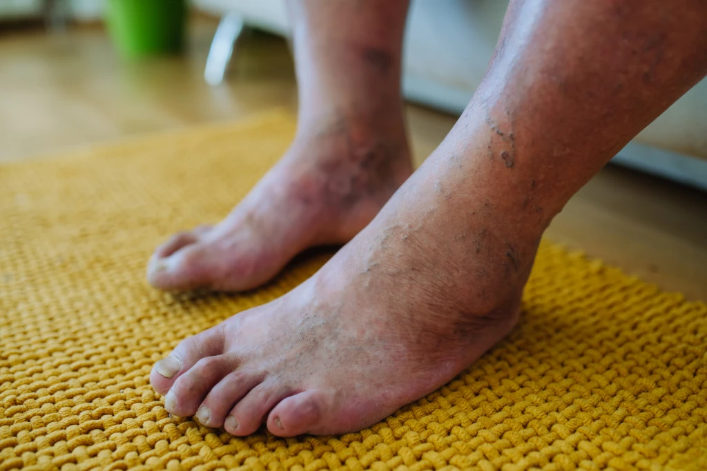
Diabetic foot is the umbrella term for the range of foot problems that diabetes can cause over time. In Singapore, where the National Population Health Survey 2024 reports that 9.1% of residents aged 18 to 74 have diabetes (external link, opens in new tab), the population at risk is large and growing. The foot is often described as the canary for diabetes complications because nerve damage, circulation problems, and slow wound healing tend to show up in the feet before they become obvious elsewhere. A new numb patch on a toe, a cut that takes weeks instead of days to close, or a swelling that appears without an obvious injury can all be the first visible sign that diabetes is starting to affect the body more broadly.
Diabetic foot care is not a single specialty’s job. Blood sugar control sits with endocrinology and family medicine. Circulation problems are diagnosed and treated by vascular surgery. Podiatry sits on the foot itself: screening for early changes, treating skin, nail, and pressure problems before they become wounds, offloading ulcers when they do appear, and coordinating with the rest of the medical team. The aim of this hub is to help patients and families recognise where they sit on the spectrum and what the structured response looks like.
What Is Diabetic Foot?
Diabetic foot is not one condition but a cluster of related ones. The common thread is that all of them are downstream effects of long-standing high blood sugar on three biological systems that meet in the foot: the nerves, the blood vessels, and the skin and wound-healing machinery.
- Nerves. Long nerves are damaged first, which is why symptoms usually start in the toes and feet rather than the hands or trunk. Loss of sensation removes the warning system that would normally stop a patient from walking on a blister or a stone in the shoe.
- Blood vessels. Both small vessels (microvascular) and larger arteries (macrovascular) are affected. Reduced blood flow slows healing and starves tissues of the oxygen and nutrients they need.
- Skin and wound healing. High glucose impairs the immune cells that fight infection, and damaged small vessels mean repair is slower. Minor breaks in the skin take longer to close and are easier for infection to take hold in.
The foot is where all three systems converge: it is the furthest point from the heart, the most loaded part of the body during the day, and the most often injured by ill-fitting shoes, hot pavements, or barefoot walking. That combination is why diabetic foot problems are the leading cause of lower limb amputation in Singapore, and why preventing them is one of the most important parts of long-term diabetes care.
Symptoms Across the Diabetic Foot Spectrum
Diabetic foot symptoms vary depending on which complication is developing. Patterns to watch for include:
- Loss of sensation or numbness in the toes and feet that builds gradually over months or years
- Burning, tingling, or shooting pain in the feet, often worse at night
- Cold feet or one foot noticeably colder than the other
- Cuts, blisters, or scrapes that take much longer than usual to heal
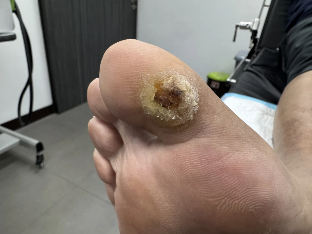
- Callus that does not shed normally, particularly if it darkens or develops a soft centre
- Swelling without an obvious injury, especially if accompanied by warmth or redness
- A visible change in foot shape, such as a collapsing arch or a rocker-bottom sole
- An open wound on the foot, with or without drainage or odour
- Pain in the calf or thigh on walking that eases with rest
Any of these in a person with diabetes is a reason to have the foot reviewed rather than waiting for the next routine appointment. The earlier the change is picked up, the more options are available to manage it.
Conditions That Make Up Diabetic Foot
Four pillar conditions sit under the diabetic foot umbrella. They overlap, they often coexist, and one can drive the next, which is why they are best understood together rather than in isolation.
- Diabetic neuropathy: nerve damage from long-standing high blood sugar. It is usually the first complication to appear, often silently, and is the underlying reason most diabetic foot injuries go unnoticed until they are advanced.
- Diabetic foot ulcer: an open wound that develops on the foot, most often on the sole, heel, or under the toes. Ulcers usually begin as something small (a blister, a callus, a tiny cut) and fail to heal because nerve damage, reduced circulation, and impaired immune response are all working against repair.
- Charcot’s foot: progressive weakening, fracture, and collapse of the bones and joints of the foot in a person whose foot has lost protective sensation. Because pain signals are blunted, the foot keeps loading through injured bone until structural deformity sets in.
- Peripheral arterial disease: narrowing of the arteries that supply blood to the legs and feet. It is closely linked to the same cardiovascular risk factors as heart disease and is a major contributor to non-healing wounds and amputation risk.
In practice, a patient often has more than one. Neuropathy and peripheral arterial disease commonly coexist. An ulcer that will not heal is often a wound in a neuropathic foot with reduced blood supply. A Charcot foot is almost always a foot with established neuropathy. Recognising the whole spectrum, not just the symptom in front of the patient, is how complications are caught earlier.
Causes of Diabetic Foot Damage
What Drives the Damage?
Diabetic foot is the cumulative effect of several diabetes-related processes playing out over years.
- Chronic hyperglycaemia. Persistently high blood sugar damages proteins in nerves and blood vessel walls, drives inflammation, and impairs cell repair.
- Microvascular damage. The smallest blood vessels in the foot are affected first, which reduces the local blood supply to nerves and skin even before the larger arteries narrow.
- Macrovascular damage. Larger arteries develop atherosclerotic plaque from the same processes that drive heart disease, narrowing the channel and reducing blood flow to the lower leg and foot.
- Neuropathy. Once protective sensation is lost, small injuries that would normally be felt and corrected go unnoticed. Pressure, friction, and minor trauma accumulate.
- Immune dysfunction. High glucose blunts the function of the immune cells that would normally clear bacteria from a small cut, so minor wounds are more likely to become infected.
These mechanisms work together rather than in isolation. A single small injury in a diabetic foot is exposed to all of them at once, which is why a problem that would resolve quickly in a healthy foot can spiral in a diabetic foot.
Who Carries a Higher Baseline Risk?
- People with long-standing diabetes, particularly with HbA1c that has been above target for years
- People with confirmed diabetic peripheral neuropathy or peripheral arterial disease
- People who have already had a foot ulcer or amputation, even if the wound has healed
- People with foot deformities such as bunions, hammertoes, or post-Charcot foot shape
- People who smoke or have smoked
- People with chronic kidney disease related to diabetes
- People who walk barefoot at home or wear ill-fitting footwear
- People with reduced vision or limited mobility that makes daily foot self-checks difficult
Diagnosis and Screening
Working out where a patient sits on the diabetic foot spectrum is a clinical assessment, not a single test. A typical podiatry review includes:
- Patient interview. Diabetes type, duration, recent HbA1c, other complications, footwear habits, daily activity, any history of foot wounds or surgery.
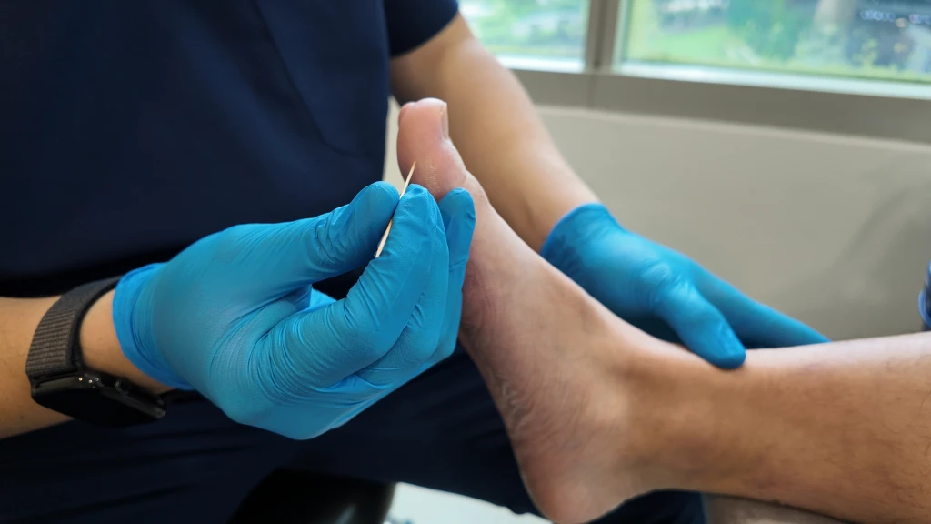
- Neurological examination. Monofilament testing to map areas where protective sensation has been lost, vibration testing with a tuning fork, and where indicated, reflex testing. This is the most sensitive way to pick up early neuropathy.
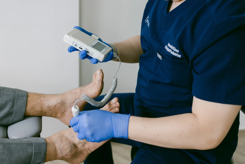
- Vascular examination. Palpation of the foot pulses, observation of skin colour and temperature, and where indicated, Ankle-Brachial Index (ABI) measurement to quantify how much blood flow is reaching the foot.
- Skin and nail check. Looking for callus, fissures, fungal infection, ingrown nails, blisters, and small wounds the patient may not have noticed.
- Foot shape and pressure assessment. Observation of arch shape, toe alignment, and any deformity, with attention to pressure points that the current footwear is not protecting.
For patients with diabetes who have not been formally screened, the structured annual review is the diabetic foot screening. The screening risk-classifies the foot (low, moderate, high, or active problem) and sets a review interval matched to that risk. International diabetic foot guidelines recommend at least one screening a year for everyone with diabetes, with shorter intervals as risk rises.
Treatment Options for Diabetic Foot in Singapore
There is no single treatment for diabetic foot because there is no single condition. Care is matched to where the patient sits on the spectrum, and most patients are co-managed across more than one specialty.
Conservative Management
The day-to-day foundation of diabetic foot care.
- Blood sugar control. Led by the diabetes team. Slowing the underlying nerve and vascular damage is the single most important factor in reducing long-term foot risk.
- Daily foot self-inspection. A short check at the end of each day for cuts, blisters, redness, swelling, or colour change. A mirror or a family member can help where reaching the soles is difficult.
- Footwear that fits. Enclosed shoes with adequate room at the toes, soft seams, and supportive soles. Avoid barefoot walking at home, particularly on tiled or rough surfaces. Replace shoes once the sole or inner lining has worn down.
- Skin and nail care. Treat dry skin, fissures, callus, and nail problems before they break the skin. In a diabetic foot, these are not cosmetic concerns.
Targeted Therapies
When a specific complication is active, treatment is matched to it.
- Offloading for ulcers. A diabetic foot ulcer cannot close if it is repeatedly compressed under body weight. Felt padding, custom orthotics, total contact casting, or a CAM walker boot redistributes pressure away from the wound so healing tissue is not crushed with every step.
- Wound debridement. Removing dead or unhealthy tissue from the wound bed so that healthy tissue can grow in. Done at intervals matched to how the wound is progressing.
- Infection management. When a wound is suspected of being infected, antibiotics are prescribed by the medical team. Dressings and wound monitoring are adjusted to track response.
- Offloading and bracing for Charcot foot. In the acute phase, full offloading (often in a total contact cast) is the priority to protect the foot while the bone consolidates. Custom footwear or bracing is built once the foot has cooled and stabilised.
- Vascular referral for peripheral arterial disease. Where reduced blood flow is limiting healing or causing rest pain, vascular surgery leads on angioplasty, stenting, or bypass. Podiatry continues local foot care alongside the vascular team.
Surgical Intervention
Surgery has a smaller role in diabetic foot than most patients expect. Most diabetic foot problems are managed conservatively. Surgical intervention is reserved for situations that conservative care cannot resolve, including vascular reconstruction for advanced peripheral arterial disease, structural correction in severe Charcot deformity, and surgical debridement or amputation when an infection cannot otherwise be controlled. These decisions sit with vascular surgery and orthopaedic surgery, with podiatry providing pre-operative and post-operative foot care.
Have Your Diabetic Foot Managed at Straits Podiatry
Diabetic foot care works best when the foot is reviewed structurally rather than only when something is already wrong. At Straits Podiatry, the assessment starts with a full review of diabetes history, current foot condition, footwear, and daily activity, so the foot’s individual risk level is clear before any plan is set.
Services that may form part of your care include diabetic foot screening with risk-matched review intervals, wound debridement and dressing management for ulcers, custom orthotic insoles and offloading footwear for high-pressure areas, skin and nail care to prevent small problems becoming wounds, and coordinated referral to endocrinology and vascular surgery within the Healthway Medical Group network where the clinical picture calls for it. Speak with the team or book a consultation to have your diabetic feet assessed and a tailored review schedule set.
Frequently Asked Questions About Diabetic Foot
How often should I have my diabetic feet screened?
International diabetic foot guidelines recommend at least one screening a year for everyone with diabetes, even if the feet currently feel fine. Patients with confirmed neuropathy, reduced circulation, a foot deformity, or a previous ulcer are screened more frequently, usually every three to six months, because the risk of a new problem is higher. Straits Podiatry sets the review interval based on a structured risk classification rather than a one-size-fits-all schedule, so your specific interval may be more or less frequent than the annual baseline.
Can diabetic foot damage be reversed?
Most of the structural changes of diabetic foot are slow to reverse, and some are not reversible. Established neuropathy usually does not return to normal even with excellent blood sugar control, although progression can be slowed substantially. Reduced circulation can sometimes be improved by vascular procedures. Calluses, skin breakdown, and early ulcers can heal with the right care. Foot shape changes from Charcot foot are typically permanent once the bone has consolidated in its new position. The realistic goal is to slow progression, protect the foot from further damage, and treat each problem as early as possible rather than expecting a full reversal.
What is the difference between diabetic neuropathy and peripheral arterial disease?
Diabetic neuropathy is nerve damage. Peripheral arterial disease is blood vessel narrowing. Neuropathy usually shows up as numbness, tingling, burning pain, or loss of sensation, often without obvious changes in how the foot looks. Peripheral arterial disease usually shows up as cold feet, pale or dusky skin, cramping leg pain on walking, slow-growing toenails, or non-healing wounds. Many patients with long-standing diabetes have both, which is why both nerve and vascular assessment are part of a proper diabetic foot screening rather than only one or the other.
When should I worry about a cut on my diabetic foot?
A cut on a diabetic foot deserves earlier attention than the same cut on a non-diabetic foot. Have any new wound reviewed within days rather than weeks, particularly if it is on the sole or heel, if it is not visibly closing within a week, if the surrounding skin is red, warm, or swollen, if there is any discharge or odour, or if you feel generally unwell. The absence of pain does not mean the cut is minor, because loss of sensation is part of the condition. The safer default is to have it checked.
What does “high-risk diabetic foot” mean?
“High-risk” is a clinical classification from international diabetic foot guidelines, not a vague description. A foot is considered high-risk when it has lost protective sensation from neuropathy, when circulation is reduced, when there is a structural deformity that creates a high-pressure area, or when there has been a previous ulcer or amputation. High-risk feet need more frequent screening, more careful footwear choice, and a lower threshold for prompt review when something changes. A patient being told their foot is high-risk is not a warning that an amputation is imminent. It is a statement that the foot needs a more structured ongoing review than a low-risk foot would.