
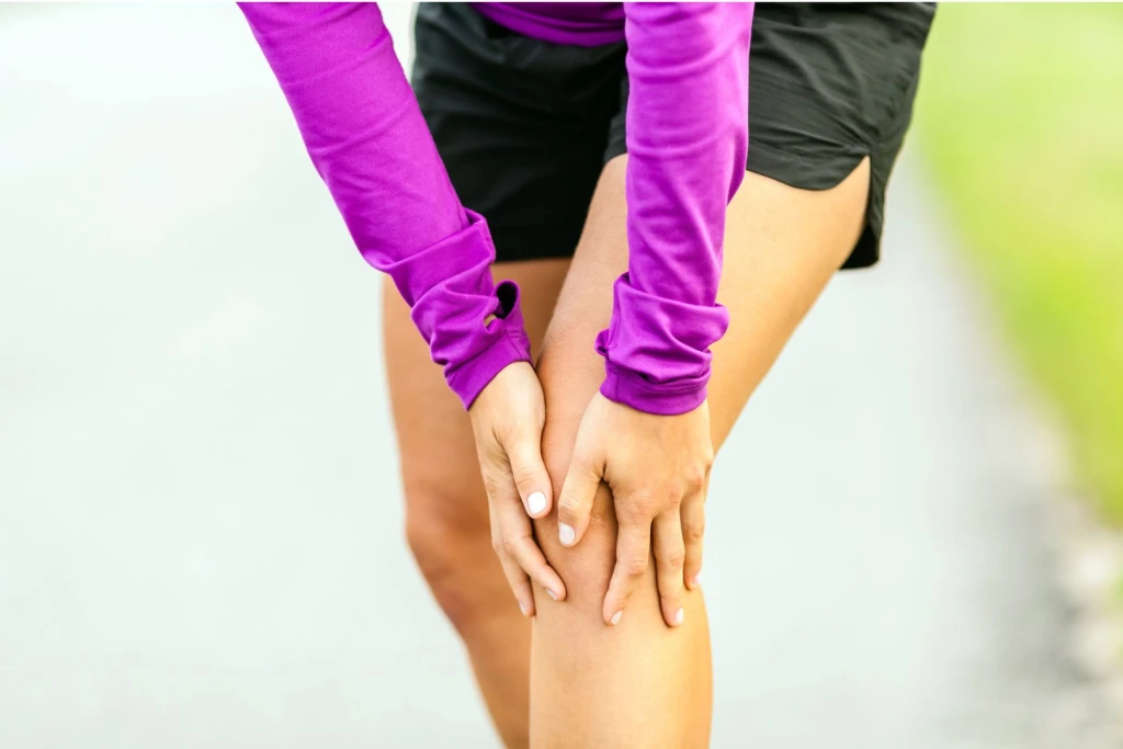
You are watching your daughter play netball at a school hall in the east, and she plants her foot to change direction when her knee buckles and she goes down clutching the front of it. The kneecap has visibly shifted to the outer side of the knee for a moment, then slid back as she straightened the leg. Or the story is quieter: an active young woman who finds her kneecap feels like it wants to slip whenever she pivots, so she has started avoiding stairs, squats, and quick turns without quite knowing why. Both pictures point to the same underlying problem.
The kneecap (patella) normally glides up and down in a shallow groove at the end of the thighbone as the knee bends and straightens. A dislocation happens when the kneecap is forced out of that groove, almost always to the outer (lateral) side, either from a direct knock or from a twisting, pivoting movement. Patellar instability is the broader pattern of a kneecap that slips, partly shifts, or feels like it will give way, whether or not it fully dislocates each time. Getting the diagnosis right matters because a first-time dislocation is an acute injury that needs medical assessment to rule out a knock-off fragment of bone or cartilage, while recurrent instability is a mechanical problem that is usually worked on conservatively before surgery is ever considered.
Symptoms of Kneecap Dislocation and Patellar Instability
An acute dislocation is usually obvious in the moment, but the instability that follows can be subtler. These are the patterns that point toward the kneecap as the source.
- A visible or felt sideways shift of the kneecap: during an episode the patella moves to the outer side of the knee. In a full dislocation it stays out until the leg is straightened, which often pops it back into place.
- The knee giving way or buckling: the leg suddenly feels like it cannot be trusted, particularly when turning, pivoting, or coming down stairs.
- Apprehension: a distinct fear that the kneecap is about to slip, which makes people guard the knee and avoid the movements that provoke it.
- Swelling after an episode: the knee often swells within hours of a dislocation, as the tissues on the inner side of the kneecap are stretched or torn as it slips out.
- Pain around the front and inner border of the kneecap: tenderness is common along the inner edge of the patella, where the stabilising ligament attaches.
- A pop or clunk sensation: patients frequently describe a pop as the kneecap leaves the groove and a second clunk as it relocates.
- Recurrent episodes with the same movements: once instability sets in, twisting, jumping, and quick direction changes tend to reproduce the slipping feeling.
Causes of Kneecap Dislocation and Patellar Instability
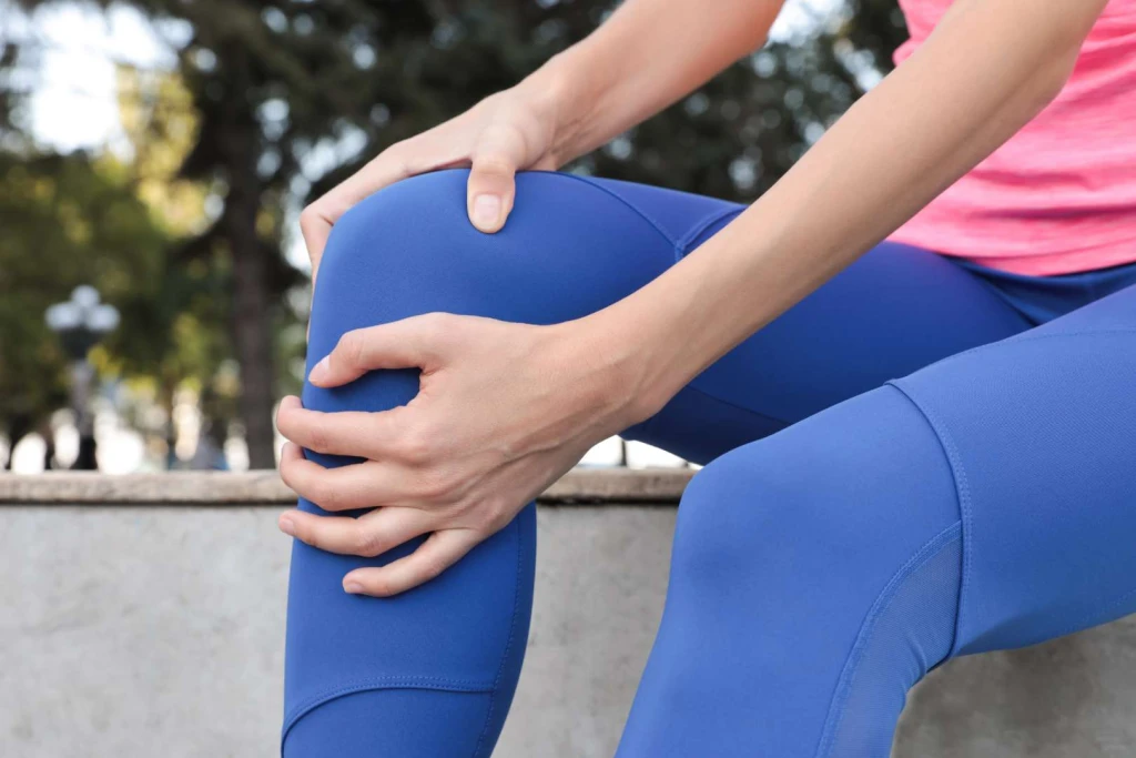
The kneecap stays centred through a balance of bony shape, soft-tissue restraints, and muscle control. Instability appears when one or more of these is not doing its job, and a first dislocation often damages the very ligament that was meant to prevent the next one.
What Causes Kneecap Dislocation and Patellar Instability?
- A twisting or pivoting injury: the most common trigger we hear about is a planted foot with a sudden change of direction, where the thigh rotates over a fixed lower leg and levers the kneecap outward.
- A direct blow to the knee: a knock to the inner side of the kneecap can push it out of its groove, common in contact sport and falls.
- Injury to the medial patellofemoral ligament (MPFL): this ligament is the main soft-tissue check that holds the kneecap on the inner side. A first dislocation usually stretches or tears it, which is why the risk of a repeat episode rises sharply afterwards.
- A shallow trochlear groove (trochlear dysplasia): when the groove the kneecap sits in is flatter than usual, there is less bony containment and the patella slips more easily.
- A high-riding kneecap (patella alta): a patella that sits high engages the groove later in the range of movement, leaving it less supported through the vulnerable early bend.
- A high Q-angle and knock-knee alignment: a wider pull-line on the kneecap, influenced by hip and knee alignment, biases it toward the outer side.
- Generalised joint hypermobility: naturally lax ligaments give the kneecap more freedom to move than is helpful.
- Weak or poorly timed quadriceps and hip muscles: the inner quadriceps (vastus medialis) and the hip muscles that control leg alignment help centre the kneecap. When they are weak or slow to fire, tracking drifts outward.
- Foot mechanics that rotate the leg inward: feet that roll in heavily (excessive pronation) increase inward rotation of the shin and dynamic knee valgus, which shifts the pull on the kneecap toward the outer side during running and landing.
Who Carries a Higher Baseline Risk?
- Adolescents and young adults, with the first dislocation frequently occurring in the teenage years.
- Young women, in whom hip width, alignment, and ligament laxity more often combine to raise the risk.
- Anyone who has already had one dislocation, because recurrence is common once the MPFL is compromised.
- People with generalised hypermobility or a family history of unstable kneecaps.
- People with knock knees, wide hips, or a naturally high-riding kneecap.
- Athletes in cutting, pivoting, and jumping sports such as netball, basketball, football, and dance.
Conditions Commonly Mistaken for Kneecap Dislocation and Patellar Instability
Not every front-of-knee problem or giving-way sensation is a patellar instability. Several other conditions produce overlapping symptoms, and separating them matters because the rehab focus and the referral pathway differ. A clinical assessment confirms which structure is actually involved.
Runner’s knee
Runner’s knee, or patellofemoral pain syndrome, produces a diffuse ache around or behind the kneecap from repeated bent-knee loading, and it can involve a mild sense of the knee giving way on stairs. The difference is that runner’s knee does not feature a kneecap that visibly slips or dislocates. The pain is one of load and tracking rather than of frank instability, though the two can coexist when tracking is poor.
Chondromalacia patellae
Chondromalacia patellae is a softening and wear of the cartilage on the underside of the kneecap, causing front-of-knee pain, grinding, and stiffness after sitting. It shares the poor-tracking background of instability and can develop alongside it, but the main complaint is cartilage-related pain rather than the kneecap leaving its groove.
Meniscus tear
A meniscus tear can also make the knee lock, catch, or give way, which is easily confused with instability. The distinction is the location and mechanism. Meniscal symptoms sit deeper in the joint line and are provoked by squatting and twisting under load, whereas patellar instability is felt at the kneecap itself as it shifts sideways.
Patellar tendonitis
Patellar tendonitis causes pain on the tendon just below the kneecap, aggravated by jumping and explosive loading. It is a tendon-overload problem with no slipping of the kneecap, so the focal tenderness on the tendon below the patella separates it from instability, which centres on the kneecap moving out of its groove.
Treating and Preventing Kneecap Dislocation and Patellar Instability
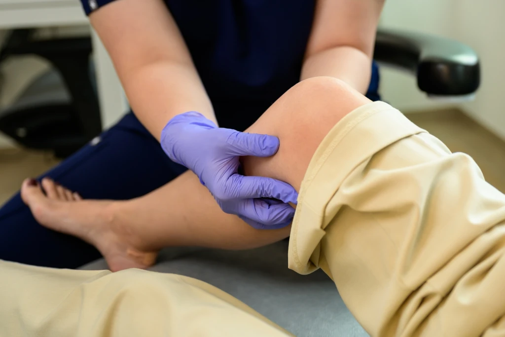
Care depends heavily on which situation you are in. A first-time acute dislocation is a medical injury: the kneecap needs to be relocated safely, and imaging is used to check for a fragment of bone or cartilage that can be knocked off as the patella slips (an osteochondral injury), because that finding changes the plan. Once the acute episode has settled, and for people whose main problem is recurrent instability, the principle is to build back the muscle control and correct the mechanical factors that let the kneecap drift outward before any surgery is considered. Structural cases, where the groove is shallow or the kneecap sits high, are the ones most likely to need an orthopaedic opinion.
Conservative treatment
For recurrent instability without a structural problem that demands surgery, these measures are usually combined and led by rehab.
- Bracing and taping: a patellar stabilising brace or a taping technique that holds the kneecap more centrally can reduce the sense of slipping during activity, particularly in the early phase while strength is being rebuilt. It is a support during rehab, not a standalone fix.
- Strengthening and control rehab: the centrepiece of conservative care. Rebuilding the inner quadriceps and the hip muscles that control leg alignment improves how the kneecap tracks and is where the durable result comes from. Our physiotherapy colleagues lead this strengthening and return-to-sport work.
- Gait analysis and biomechanical assessment: where foot and leg mechanics are feeding dynamic knee valgus, a lower-limb biomechanical review identifies the contribution. This is the part of the picture podiatry is best placed to address.
- Custom foot orthoses: where heavy pronation is rotating the shin inward and biasing the kneecap outward, orthoses can reduce that inward drive during walking, running, and landing, supporting the strengthening work rather than replacing it.
- Activity modification and graded return: stepping back from cutting, pivoting, and deep-squat loading while control is rebuilt, then reintroducing sport-specific movement gradually once the knee feels secure. Returning by staged milestones rather than by feel reduces the chance of another episode.
When conservative care isn’t enough
A first-time dislocation always warrants medical assessment on the day, both to relocate the kneecap safely if it has not already gone back and to rule out an osteochondral fragment with imaging. Beyond that, where the kneecap keeps dislocating despite a genuine course of strengthening and bracing, or where imaging shows a structural cause such as marked trochlear dysplasia, patella alta, or a torn MPFL, an orthopaedic surgeon is the right person to advise on options. These can include MRI to map the soft-tissue and cartilage damage, MPFL reconstruction to restore the inner check on the kneecap, and bony procedures such as a tibial tubercle transfer or trochleoplasty for the underlying shape problem. Podiatry and physiotherapy support the rehabilitation before and after any such surgery, but the decision to operate sits with the orthopaedic team.
Have Your Patellar Instability Assessed at Straits Podiatry
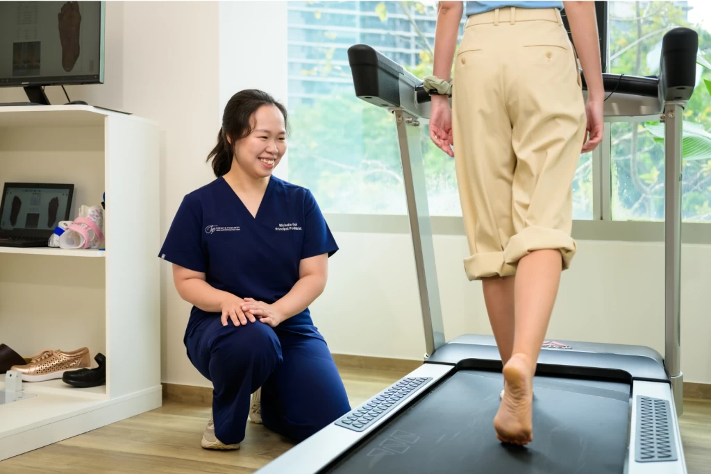
A dislocating or unstable kneecap sits across orthopaedics, physiotherapy, and podiatry, and honest care means being clear about who does what. A first-time dislocation belongs with a doctor on the day. For recurrent instability, once the acute injury is excluded, much of the durable work is muscle control and mechanics. At Straits Podiatry, an assessment for an unstable kneecap includes a gait analysis and a lower-limb biomechanical review to identify whether foot and leg mechanics are feeding the outward pull, and where they are, custom foot orthoses can help reduce it.
We work alongside our physiotherapy service on the strengthening and return-to-sport side, and we will refer you on to an orthopaedic surgeon where the picture is structural or the kneecap keeps dislocating. If your kneecap has slipped, given way, or feels like it wants to, book a consultation for an assessment at any of our three Singapore clinics.
Frequently Asked Questions About Kneecap Dislocation and Patellar Instability
What should I do if my kneecap dislocates?
Treat a first-time dislocation as an acute injury and get it assessed the same day. Often the kneecap slides back on its own as you gently straighten the leg, but even when it does, you should still be seen so a fragment of bone or cartilage can be ruled out on imaging and the knee protected while the stretched tissues settle. Do not force the kneecap back yourself. Support the leg, apply ice to control swelling, keep weight off it, and arrange to be reviewed rather than pushing through.
Will my kneecap dislocate again?
Recurrence is common after a first dislocation, because the episode usually stretches or tears the ligament (the MPFL) that holds the kneecap on the inner side. The risk is higher in younger patients, in those with a shallow groove or a high-riding kneecap, and in anyone with naturally lax ligaments. A structured course of strengthening and, where relevant, bracing and mechanical correction lowers that risk, but structural cases may keep slipping until the underlying shape is addressed surgically.
Can patellar instability be improved without surgery?
For many people, yes. When there is no major structural problem, recurrent instability responds to rebuilding the inner quadriceps and hip control, using a stabilising brace or taping during the rebuild, and correcting foot and leg mechanics that bias the kneecap outward. Surgery is generally reserved for kneecaps that keep dislocating despite genuine rehab, or for clear structural causes such as marked trochlear dysplasia or an incompetent MPFL. The assessment sorts which group you are in.
Can a podiatrist help with an unstable kneecap?
Podiatry addresses one specific part of the picture: how foot and leg mechanics load the knee. Feet that roll in heavily increase inward rotation of the shin and dynamic knee valgus, which shifts the pull on the kneecap outward, so a gait analysis and biomechanical review can identify where mechanics are contributing, and custom orthoses can reduce that drive. The muscle-control rehabilitation is led by physiotherapy, and structural cases go to an orthopaedic surgeon. Podiatry is one honest part of a coordinated plan, not the whole of it.
Do custom orthotics help patellar instability?
Orthoses help when foot mechanics are genuinely part of the problem, particularly with heavy pronation that rotates the shin inward and biases the kneecap toward the outer side. They are not a fix on their own and they do nothing for the ligament or the bony shape of the groove. Their role is to reduce one mechanical contributor so the strengthening work has a fairer chance. A biomechanical assessment shows whether orthoses are likely to make a difference for your particular knee.
How long does recovery take after a kneecap dislocation?
A first dislocation without a bone or cartilage fragment often settles enough for a graded return to activity over six to twelve weeks, depending on how much the inner tissues were injured and how consistent the rehabilitation is. Recurrent instability worked on conservatively takes a similar block of focused strengthening before the knee feels reliably secure. Where surgery is needed for a structural cause, recovery is longer and is guided by the orthopaedic team. Returning by staged milestones rather than by how the knee feels on a good day is what tends to hold the result.