

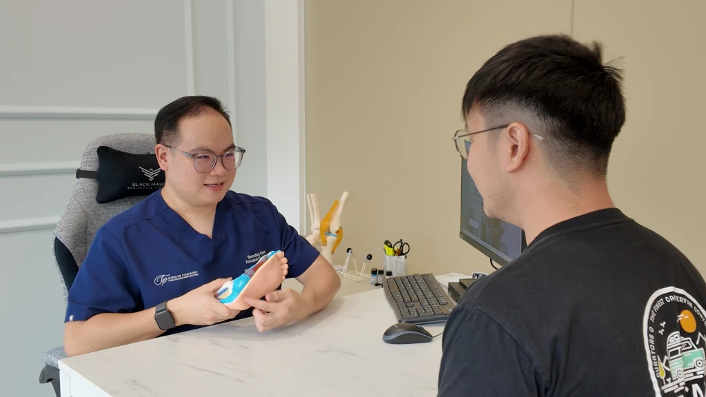
A patient walks into the consultation room, sits down, and starts describing their heel pain. By the time they reach the second sentence, a fair amount of the diagnostic work is already underway. The first 60 seconds of any heel-pain visit is densely packed with information, most of it gathered before any structured examination begins.
This blog is the inside view. What I am looking at, what I am listening for, and how those early observations shape every question and palpation that follows.
Why the First 60 Seconds Carries So Much Information
Heel pain has at least nine recognised causes, several of which produce similar pain in similar regions. The first minute is useful because the patient has not yet had time to perform their pain story. They walk the way they actually walk, and they describe the pain in their own words before clinical vocabulary takes over.
Once an examination starts, behaviour shifts. People stiffen up, brace, demonstrate. The unguarded first minute often tells me more than the next fifteen.
The Walking-Pattern Observations Before You Even Sit Down
Three things I notice in the walk from the door to the chair.
Where they offload. Patients with under-heel pain tend to land further forward on the foot, almost a forefoot-first gait. Patients with back-of-heel pain stiffen at the ankle and shorten their stride to avoid the push-off phase. A sudden-onset limp is asymmetric; a long, grumbling history walks more symmetrically because the body has had time to redistribute load.
What the shoes look like. The wear pattern under the heel, the collapse of the heel counter, whether the shoe is appropriate for the patient's daily load. A new pair of minimalist shoes worn for the past month is a useful clue. So is a five-year-old pair worn into the ground.
How they sit down. Plantar fasciitis patients often plant the painful foot first and unload it quickly. Patients with heel fat pad syndrome avoid hard heel-strike on the chair leg as they lower themselves. Patients with Achilles tendonitis often keep the heel slightly lifted as they settle.
None of these on their own is diagnostic. Together they give a working hypothesis before the conversation has formally started.
The Four Questions That Narrow the Diagnosis Fast
Once the patient is seated, four questions do most of the early diagnostic work. The order matters.
1. When is the pain at its worst, and what makes it ease? First-step pain that eases after a few minutes points toward plantar fasciitis. Pain that builds through the day without easing points toward fat pad irritation or a bony stress injury. Pain that warms up during activity and worsens after points toward Achilles involvement.
2. What does the pain feel like? Sharp and stabbing is mechanical tissue. Deep and bruised is fat pad. Burning, tingling, or electric is nerve, including Baxter's nerve entrapment. Patients use these words spontaneously if I let them, and the spontaneous ones are usually more accurate than the clinical words they reach for once prompted.
3. What changed in the four weeks before this started? New shoes, a job change involving more standing, a return to running after a layoff, a sudden increase in walking volume, a holiday with long days on hard floors. Heel pain rarely arrives without a load change behind it. The change is sometimes small, and the patient often does not connect it until I ask.
4. What has been tried, and how did the heel respond? A heel that improved with rest and flared again with return to load is behaving like a tendinopathy. A heel that did not respond to a few weeks of sensible offloading is behaving like something more structural. A heel that got worse with stretching points toward something that does not want to be stretched, which itself narrows the list.
The Palpation Order That Separates the Common Diagnoses
When I get to the foot itself, I work from the highest-probability finding outward.
Medial calcaneal tubercle first. The underside of the heel, slightly toward the inside of the foot, where the plantar fascia attaches. Sharp, reproducible tenderness here is the classic plantar fasciitis finding.
Central heel pad next. A deep, bruised quality on direct heel pressure, particularly compared to the other foot, suggests fat pad involvement. The location overlaps with plantar fasciitis but the quality of the pain is different.
Medial to the tubercle, slightly deeper. The Baxter's nerve region. Reproducing burning, electric, or radiating pain here changes the diagnostic direction entirely.
Achilles insertion and the soft spot above it. Tenderness on the bony insertion is insertional Achilles. Tenderness 2 to 6 cm above is mid-substance Achilles. Tenderness at the bony prominence behind the tendon is Haglund's. Three different conditions in a small region.
Calcaneal squeeze. Compressing the heel from both sides at once. Pain on calcaneal compression in a runner with a recent training spike is a stress-fracture flag and changes the plan immediately.
That sequence either confirms the working hypothesis or surfaces a second condition sitting alongside the first. In real clinic life, single-condition heel pain is the textbook version. Mixed presentations are more common than the textbooks suggest.
What I Look for That Imaging Cannot Show
Patients often arrive with a scan. Imaging is useful, but it has limits, and the first 60 seconds is where I notice what imaging cannot: how the foot loads dynamically, whether the calf is bearing its share of work or the foot is compensating, whether one heel is taking the majority of body weight standing still because the other knee or hip has changed the patient's posture, whether the pain has spread up the leg in a pattern that suggests the heel is no longer the only problem.
Imaging shows the static structure. The first 60 seconds shows how the patient lives in their body. Both matter, and neither replaces the other.
When the First 60 Seconds Is Not Enough
Some presentations need more than pattern matching, and self-management tends to stall on these:
- Night pain or rest pain, particularly if it wakes the patient up.
- Pain that started with a sudden pop or snap during activity.
- Pain accompanied by swelling, warmth, or systemic symptoms.
- Heel pain in a patient with diabetes, regardless of the pattern.
- Pain treated for plantar fasciitis for months without response (a frequent setup for missed Baxter's nerve entrapment or a missed stress reaction).
- Pain with no clear load story behind it, particularly in an older patient.
In these cases the assessment expands to include more careful history, a wider examination, and a lower threshold for imaging.
The first minute of an assessment is not magic. It is pattern recognition built on the same conditions everyone else sees, with attention paid to the unguarded details patients reveal before they realise they are revealing them. Heel pain can be easily misdiagnosed, which is why the unguarded first minute matters as much as the structured examination that follows.
Book a consultation at any of our three locations across West, Central, and East Singapore when you are ready.